 6.6 "SKELETON" Tissues And Supporting System
 The skeleton is the supporting framework of an organism. It is typically made out of hard, rigid tissue that supports the form of the animal’s body and protects vulnerable organs.
For land-dwelling animals, skeletons are also necessary to support movement, since walking and flying rely on the ability to exert force on rigid levers such as legs and wings.
Arthropods such as insects have an “exoskeleton” – an outer covering of a hard material called chitin that protects their internal tissues and allows them to walk, jump, and fly.
Vertebrates such as humans have internal skeletons, made of a tissue called bone that gives the limbs their rigidness and protects vital organs such as the heart and brain.
The term “vertebrate,” in fact, comes from a specific part of the internal skeleton – “vertebrae” are small bones that encase and protect the spinal cord, a vital tissue that acts as the information channel between the brain and the rest of the body.
The skeletons of most vertebrates, including humans, are made of bones. Bones are complex structures consisting of many different types of tissues, which perform both structural and biological functions.
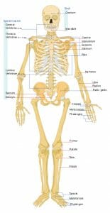
The image below shows the human skeleton with some of the most important bone groups labeled:

For anatomy students and medical students, it’s important to note that this skeleton’s right forearm is rotated forward to show how the arm bones look from a different angle. This is not the standard positioning found in most anatomy diagrams, so keep in mind that in most diagrams, both arms are positioned like this skeleton’s left arm.
Here we’ll talk more about the functions of the skeleton and the structures, functions, and classifications of its bones.
6.7 FUNCTION OF SKELETON For vertebrates such as humans, the skeleton performs many essential functions. Some are directly related to the purpose of all skeletons of providing structural support, protection, and support for locomotion. Others are biological functions unrelated to structural support that have been adopted by vertebrate bone tissues over time.
The functions of the skeleton include: 1.
Structural Support The skeleton serves the vital purpose of giving form to an animal’s body. Some animals that live in water, such as the octopus, have no skeleton. This is possible because their tissues are partially supported by the water that surrounds them, which is much heavier than air and allows some of an animal’s body structures to float. You’ll notice that octopi don’t do so well on dry land!
For land animals, it’s essential to have a skeleton that fights the force of gravity, which might otherwise prevent movement and even crush organs. That’s why all mobile land animals have either an exoskeleton, like those of insects and spiders, or an internal skeleton, like those of humans and other vertebrates.
2.
Locomotion Almost all forms of on-land locomotion require the ability to push rigid levers against our environment. When we walk, our leg bones are levers that exert force on the ground to propel us forward. When birds fly, their wing bones are levers that push against air molecules to allow them to move.
The role of bones in locomotion is the major reason why broken bones can be a death sentence for animals in the wild. Without intact levers to push against, animals can be unable to move quickly or at all, which in turn renders them unable to find food or escape from predators.
3.
Protection In addition to supporting the body’s structure against the force of gravity and allowing locomotion, the skeleton plays the vital role of protecting important organs from injury. Some important protective bones in the human body include:
The skull is a thick covering of bone that protects the brain from injury.
The spinal column – made of “vertebrae,” from which “vertebrates” get their name – protects the spinal cord, which is the major nerve cord that allows the brain to communicate with the body.
The ribcage forms a protective barrier around the lungs and heart, without which the body would not be able to supply blood to the brain and would soon die.
The vertebrate body must make compromises between protection and mobility. Our lower abdominal organs such as the intestines, for example, are not protected by the ribcage. But this lack of a hard covering around our abdomen allows us to bend climb, and shift our weight in a way that greatly enhances our mobility!
4.
Blood cell production For animals with internal skeletons, the bones also perform other vital biological functions that are not directly related to their role as structural support. In humans, one of the most important of these roles is blood cell production.
Our bones are made out of living tissue. Their outer tissues are hard and rigid, but their inner tissues are soft and serve other purposes. On the inside of our bones – in the part called the “bone marrow” – the stem cells that create our red and white blood cells can be found.
Without healthy bone marrow, our bodies would stop replacing their blood cells – and would soon lose the ability to transport oxygen and fight infection!
This is why bone marrow transplants are sometimes prescribed for people with “blood cancers.” In blood cancers such as leukemia, the cancerous cells actually originate in the bone marrow. These cancerous cells produce large numbers of blood cells, but the blood cells they produce do not work properly.
As a result, people with cancer of the stem cells that produce white blood cells may have very high white blood cell counts – but they have difficulty fighting infection, because these white blood cells produced by cancerous stem cells do not work properly.
In bone marrow transplants, doctors attempt to kill off the stem cells in a patient’s own bone marrow, and then replace some of the marrow with marrow from a healthy donor that can produce healthy blood cells.
5.
Storage Bones can store calorie-rich fat and minerals that other body tissues might need at a later date.
The hard part of bone tissue is rich in calcium, which in emergencies the body can release from the bones to serve other purposes.
Yellow bone marrow tissue is composed primarily of fat, which can act as a storage point for calories and nutrients.
Red bone marrow tissue is rich in iron, a necessary ingredient for red blood cells. Iron deficiency is a common cause of anemia – a condition in which insufficient production of red blood cells can lead to weakness, fatigue, dizziness, and even fainting.
6.
Endocrine regulation Bone cells release a hormone called osteocalcin, which has effects on blood sugar, fat storage, and male sex hormones.
The release of osteocalcin by bone cells prompts the pancreas to release more insulin, resulting in lowered blood sugar and increased consumption of sugar by cells. It also causes fat cells to release a hormone called adiponectin, which prompts the breakdown of fat for energy.
Osteocalcin directs the male testes to produce more testosterone, and is also thought to encourage the body to produce more bone cells.
The complex interplay between hormones in the human body is not well-understood. In this case, it’s possible that by prompting the release of insulin and the breakdown of fats for energy, it may be freeing up additional energy that the body can use to grow more bone cells.
6.8 TYPES OF BONE While all bones are made of similar tissue, there are a few different kinds of bones that have different characteristics and growth patterns, which allow them to serve their different functions in the human body. These are:
1.
LONG BONES Long bones are those that play a vital role in locomotion and in supporting our weight against the force of gravity. These include the long bones of the arms, legs, hands, and feet.
Long bones lengthen substantially as a person grows, and have a “growth plate” or “epiphyseal plate” at their ends, where new bone is formed during growth.
In children, most blood cells are produced by red bone marrow in the long bones. In adults, much of the red marrow in long bones is replaced by yellow marrow, and blood cell production takes place mostly in the flat bones.
Doctors can sometimes tell a person’s approximate age from looking at their epiphyseal plates, as these shrink and change as a person ages and stops growing. This approach is sometimes used to estimate the age of a person or animal at time of death by forensic investigators, archaeologists, and paleontologists.
2.
SHORT BONES Short bones are roughly cube-shaped bones that offer support and mobility in complex structures like the wrist and ankles.
Both our wrists and ankles require a complex large range of motion – but also require extreme strength and stability, especially our ankles which must support our weight.
To solve this problem, the body uses a series of interlocking cube-shaped bones, held together by strong ligaments. These provide a solid structure, but can also be shifted against each other to produce large or small changes in the shape and position of our hands and feet.
3.
FLAT BONES Flat bones serve the primary purpose of protecting important organs. These bones to not require the same range of motion like the short bones, and to not undergo pronounced growth like the long bones.
In adults, most blood cell production occurs in the red bone marrow of the flat bones.
Examples of the flat bones include the skull, sternum, ribs, and the scapulae, which protect our lungs and heart from the back.
4.
IRREGULAR BONES Irregular bones are bones that have complex shapes, which allow them to serve highly specific purposes. They often serve to both protect internal organs and give structure to the body.
Examples of irregular bones include the vertebrae themselves, whose complex shape allows them to shield the spinal column from all sides while allowing our spines to be mobile and flexible.
The pelvic girdle, which protects some internal organs while also providing a structural base for our legs, is another example of an irregular bone.
5.
SESAMOID BONES Sesamoid bones are bones that are embedded in tendons, and which provide additional shielding and cushioning for high-stress, high motion areas of the body.
One example of a sesamoid bones is the patella – a small, round bone which covers the kneecap and protects the tendons underneath. Sesamoid bones are also found in the hands, knees, and feet.
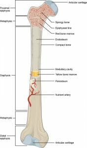
The structure of bones is best exemplified by looking at long bones, which undergo the most growth and which contain distinct cavities for bone marrow. Long bones contain several types of tissues, each of which assist with the functions our bones must perform.
6.9 THE BONE STRUCTURE COMPACT BONE Compact bone
, also called “
cortical bone,” is the hard outer shell of all bones. It consists of “
osseous tissue” made of “
osteocytes,” or bone cells.
In osseous tissue, bone cells are surrounded by a solid matrix of minerals and proteins. The most important mineral is hydroxyapatite, a calcium- and phosphorous-rich mineral also found in tooth enamel and in some naturally-occurring rocks.
Hydroxyapatite is hard and solid, but also prone to shattering, so it is interlaced in compact bone tissue with collagen fibers. Collagen is same tough, strong type of protein found in skin. The result is a matrix that is hard and solid, but also has flexibility and resilience.
The basic structural units of cortical bones are “osteons” – microscopic cylinders of bone tissue. Through the center of each cylinder runs a cord of blood vessels and nerves.
This structure of tiny cylinders ensures that bone cells receive the oxygen and nutrients they need to survive.
SPONGY BONE Spongy bone, also known as “
cancellous bone,” is bone that has a “spongy” structure that consists of fibers of hard bone tissue interlaced with softer tissues such as blood vessels and bone marrow.
Spongy bone is structurally weaker than more dense types of bone tissues, but provides an excellent site for important biological functions such as the exchange of calcium ions with the blood and the production of red blood cells.
Spongy bone is found near the ends of long bones, and inside of vertebrae.
ARTICULAR CARTILAGE Articular cartilage is, as the name suggests, cartilage – the same tough but flexible substance that makes up our noses and ears. At the ends of bones, cartilage provides cushioning and flexibility, allowing bones to slide past each other and permitting movement.
The name of “articular cartilage” comes from the verb “articulate,” which means “to unite by a joint.” This means that two structures are connected, but are able to move relative to each other because of the joint between them.
Pain and injury can result when this articular is torn or worn away, causing the hard, rigid surfaces of bones to rub against each other directly. Torn cartilage in the knee is one common sports injury that can occur when the knee joint is hit hard or wrenched violently.
In some cases, surgical repair of the cartilage is necessary to avoid damage to the hard outer shells of leg bones from rubbing against each other directly.
EPIPHYSEAL PLATE As mentioned above, “epiphyseal” refers to the region of bone where the bone can grow by producing new bone cells.
In children and teens, an “epiphyseal plate” is a site of active growth and new bone production; in adults, must of this growth tissue disappears, and what is left is an “epiphyseal line” that shows where growth once occurred. These epiphyseal regions are most obvious at the ends of long bones, which undergo extensive growth during a person’s lifetime.
The epiphyseal plate is made of cartilage – that same tough-but-flexible substance we heard about earlier. Bones grow by laying down a layer of cartilage, and then using that cartilage as a matrix for the growth of new bone cells. In that way new bone growth starts out flexible but grows strong and solid, and the cartilage helps to direct the shape and growth patterns of the new bone cells.
Soft areas that allow bone growth can also be found in other types of bone, such as the “fontanelle” of the skull. The fontanelle is the “soft spot” on a baby’s head, where soft tissue has not yet been replaced by bone cells because the skull still needs to have flexibility to accommodate the growing brain.
In adults the fontanelle, like the epiphyseal plate, disappears, leaving only a trace of its existence in the form of the seam where the bone plates of the skull fused.
RED BONE MARROW Red bone marrow, unsurprisingly, is where red blood cells are made. It is also the site of production of white blood cells and platelets.
At birth, most bone marrow in the body is red marrow; as a person ages, about half of that red marrow is converted into yellow marrow, leaving red marrow mostly near the ends of long bones and inside the flat bones such as the pelvis and sternum.
This iron-rich tissue is vital to the health of the entire body as it is responsible for producing the vehicles that carry oxygen to the brain and other organs, and the cells that prevent and fight infection.
Diseases that destroy red bone marrow or leave it unable to function can lead to severe complications, including death.
YELLOW BONE BARROW Yellow bone marrow is a fatty tissue which contains stem cells that produce fat, cartilage, and bone. It does not produce blood cells, but rather functions mainly to store fat and provide the right environment to keep the surrounding bone healthy.
In cases of extreme blood loss or illness, yellow marrow can convert back into red marrow to help replace lost blood cells.
PERIOSTEUM The periosteum is the membrane that covers the outer surface of bones. The only exception is the joints of the long bones, which are covered by cartilage cushioning.
The periosteum is made of connective tissue rich in collagen. It also contains nerve endings that can feel pain. This allows the periosteum to serve the double purpose of protecting our bones by resisting trauma, and informing us through pain when something is wrong.
The periosteum is also the site of new bone growth by bone cells called “osteoblasts” as bones grown and become thicker.
Even as new bone is laid down by osteoblasts beneath the periosteum, cells called “osteoclasts” digest bone tissue from the inside, widening the bone’s internal marrow-containing cavity and preventing the bone shell from becoming too thick.
NUTRIENT ARTERY A nutrient artery is a part of the circulatory system responsible for delivering oxygen, nutrients, and other vital materials to the bone.
These arteries enter the bones through canals called “foramina,” which are holes in the bone that exist for the purpose of allowing arteries to enter the bone tissue.
From the nutrient artery, blood vessels branch down to the level of the osteons, where microscopic blood vessels run through the center of tiny cylinders of bone cells.
If nutrient arteries become blocked or damaged, bone tissue death and infection can result. Although bones may look like solid, “dead” tissue, they require the activities of living cells to stay strong and to avoid becoming prey for dangerous pathogens.
ENDOSTEUM The endosteum is the membrane that covers the inside of the bone’s medullary cavities – the hollow spaces that are typically filled with marrow.
The endosteum greatly resembles the periosteum, consisting of a thin layer of very tough fibrous tissue, which also contains nerve cells.
During periods of starvation, the body can absorb and digest the endosteum along with some of the fat from bone marrow. This results in a weakening of the bones, but can help the body continue to function for longer until nutrients can be found.
The endosteum is also the site of bone reabsorption as bones grow and become thicker. To prevent the hard shell of compact bone from becoming too thick, bone tissue is absorbed and digested by cells called “osteoclasts” on the inside of the medullary cavity at the same time that new layers are being laid down on the outside of the bone, under the periosteum.
This ingenious mechanism allows bones to grow in both length and thickness while expanding the medullary cavity in tandem with the growth of the compact bone tissue.
Scroll Down & Continue Reading!


